CYBERMED LIFE - ORGANIC  & NATURAL LIVING

Ultrasound therapy

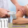
Ultrasound therapy: Therapeutic ultrasound refers generally to any type of ultrasonic procedure that uses ultrasound for therapeutic benefit. This includes HIFU, lithotripsy, targeted ultrasound drug delivery, trans-dermal ultrasound drug delivery, ultrasound hemostasis, cancer therapy, and ultrasound assisted thrombolysis  It may use focused ultrasound (FUS) or unfocused ultrasound.

Ultrasound is a method of stimulating the tissue beneath the skin's surface using very high frequency sound waves, between 800,000 Hz and 2,000,000 Hz, which cannot be heard by humans.

There is little evidence that active ultrasound is more effective than placebo treatment for treating patients with pain or a range of musculoskeletal injuries, or for promoting soft tissue healing.

Relatively high power ultrasound can break up stony deposits or tissue, accelerate the effect of drugs in a targeted area, assist in the measurement of the elastic properties of tissue, and can be used to sort cells or small particles for research.

  • Focused high-energy ultrasound pulses can be used to break calculi such as kidney stones and gallstones into fragments small enough to be passed from the body without undue difficulty, a process known as lithotripsy.
  • Cleaning teeth in dental hygiene.
  • Focused ultrasound sources may be used for cataract treatment by phacoemulsification.
  • Ultrasound can ablate tumors or other tissue non-invasively. This is accomplished using a technique known as High Intensity Focused Ultrasound (HIFU), also called focused ultrasound surgery (FUS surgery). This procedure uses generally lower frequencies than medical diagnostic ultrasound (250–2000 kHz), but significantly higher time-averaged intensities. The treatment is often guided by Magnetic Resonance Imaging (MRI); the combination is then referred to as Magnetic resonance-guided focused ultrasound (MRgFUS).
  • Delivering chemotherapy to brain cancer cells and various drugs to other tissues is called acoustic targeted drug delivery (ATDD). These procedures generally use high frequency ultrasound (1–10 MHz) and a range of intensities (0–20 W/cm2). The acoustic energy is focused on the tissue of interest to agitate its matrix and make it more permeable for therapeutic drugs.
  • Ultrasound has been used to trigger the release of anti-cancer drugs from delivery vectors including liposomes, polymeric microspheres and self-assembled polymeric.
  • Ultrasound is essential to the procedures of ultrasound-guided sclerotherapy and endovenous laser treatment for the non-surgical treatment of varicose veins.
  • Ultrasound-assisted lipectomy is Liposuction assisted by ultrasound.

There are three potential effects of ultrasound. The first is the increase in blood flow in the treated area. The second is the decrease in pain from the reduction of swelling and edema. The third is the gentle massage of muscle tendons and/ or ligaments in the treated area because no strain is added and any scar tissue is softened. These three benefits are achieved by two main effects of therapeutic ultrasound. The two types of effects are: thermal and non thermal effects. Thermal effects are due to the absorption of the sound waves. Non thermal effects are from cavitation, microstreaming and acoustic streaming.

Cavitational effects result from the vibration of the tissue causing microscopic bubbles to form, which transmit the vibrations in a way that directly stimulates cell membranes. This physical stimulation appears to enhance the cell-repair effects of the inflammatory response.

  • A study to evaluate the effect of ultrasound treatment on nodules in multiple sclerosis patients.

    facebook Share on Facebook
    Abstract Title:

    A study to evaluate the effect of ultrasound treatment on nodules in multiple sclerosis patients.

    Abstract Source:

    Int J Neurosci. 2016 May 4:1-20. Epub 2016 May 4. PMID: 27144498

    Abstract Author(s):

    Anna Gil Sánchez, Eugenia Lacasa Andrade, Joan Valls Marsal, Lourdes Assens Tauste, Cristina González Mingot, Jorge Lecina Monge, Margarita Casalilla Puértolas, Nuria Sáez Calero, Albert Sacristán Argemí, Blanca Guiu Badia, Silvia Peralta Moncusí, Hugo Gonzalo Benito, Lara Nogueras Peñabad, Marta Lordan Pujol, Mónica Nieves I Collado, Luis Brieva Ruiz

    Article Affiliation:

    Anna Gil Sánchez

    Abstract:

    :Purpose/Aim: Ultrasound has demonstrated anti-inflammatory and pain-relief benefits in several conditions such as cellulite or trauma events. We assessed the efficacy of ultrasound therapy on nodules associated with first-line treatments in multiple sclerosis patients.

    MATERIALS AND METHODS:Twenty-two multiple sclerosis patients were enrolled during 2013 and randomized to two groups: in the control group patients were treated only with a conventional gel prescribed for cellulite and nodules, while in the experimental group the gel was combined with ultrasound therapy. Patients were treated during 10 weeks and followed up for 10 additional weeks. Three nodules were assessed for each patient, measuring size, pain and redness at 0, 10 and 20 weeks.

    RESULTS:We found a significant decrease in both groups in size, pain and redness across the three visits (p<0.0001 for size, p = 0.01 and p<0.0001 for pain, and p = 0.0002 and p<0.0001 for redness, respectively for the difference at visit 2 and 3 with respect to visit 1). More interestingly, we observed a greater reduction in pain and redness in the ultrasound-treated group, but the difference was only statistically significant at 10 weeks (p = 0.01 for both pain and redness). On the third visit, no differences between control and experimental groups were detected, both achieving the same levels in measured variables.

    CONCLUSIONS:Both treatments are useful to improve skin reaction after first-line treatments, but ultrasound in combination with gel achieves a faster reduction in pain and redness, suggesting that ultrasound treatment might be a good analgesic for nodule management in multiple sclerosis patients.

  • Case report: A breast cancer patient treated with GcMAF, sonodynamic therapy and hormone therapy. 📎

    facebook Share on Facebook
    Abstract Title:

    Case report: A breast cancer patient treated with GcMAF, sonodynamic therapy and hormone therapy.

    Abstract Source:

    Anticancer Res. 2014 Aug ;34(8):4589-93. PMID: 25075104

    Abstract Author(s):

    Toshio Inui, Kaori Makita, Hirona Miura, Akiko Matsuda, Daisuke Kuchiike, Kentaro Kubo, Martin Mette, Yoshihiro Uto, Takahito Nishikata, Hitoshi Hori, Norihiro Sakamoto

    Article Affiliation:

    Toshio Inui

    Abstract:

    Gc protein-derived macrophage-activating factor (GcMAF) occurs naturally in the human body. It has various functions, such as macrophage activation and antitumor activities. Recently, immunotherapy has become an attractive new strategy in the treatment of cancer. GcMAF-based immunotherapy can be combined with many other therapies. Sonodynamic therapy (SDT) using low-intensity ultrasound is a novel therapeutic modality. Ultrasound has been demonstrated to activate a number of sonosensitive agents allowing for the possibility of non-invasive targeted treatment for both superficial and deep-seated tumors. The current case study demonstrates that GcMAF and SDT can be used in combination with conventional therapies in patients with metastatic cancer, especially where treatment options are limited due to factors such as toxicity. This case study also suggests a new concept of cancer treatment using local destruction of cancer tissue, in this case conducted with SDT, to be used in combination with GcMAF immunotherapy as a systemic treatment.

  • Cesarean scar pregnancy: noninvasive and effective treatment with high-intensity focused ultrasound.

    facebook Share on Facebook
    Abstract Title:

    Cesarean scar pregnancy: noninvasive and effective treatment with high-intensity focused ultrasound.

    Abstract Source:

    Am J Obstet Gynecol. 2014 Oct ;211(4):356.e1-7. Epub 2014 Apr 23. PMID: 24769010

    Abstract Author(s):

    Juhua Xiao, Shouhua Zhang, Fang Wang, Yuqin Wang, Zhen Shi, Xin Zhou, Jinshui Zhou, Jinshi Huang

    Article Affiliation:

    Juhua Xiao

    Abstract:

    OBJECTIVE:The aim of this preliminary study was to investigate whether ultrasound-guided high-intensity focused ultrasound (HIFU) can play a role in treating cesarean scar pregnancy (CSP).

    STUDY DESIGN:Between November 2011 and December 2012, 16 patients with CSP were treated with ultrasound-guided HIFU ablation. Successful treatment was defined as disappearance of CSP mass, undetectable serum beta human chorionic gonadotropin, and no serious complications such as severe bleeding, uterine rupture, or hysterectomy.

    RESULTS:All patients were successfully treated in the outpatient department and none required readmission. After 2-5 treatment sessions, the mean time for achieving undetectable serum beta human chorionic gonadotropin was 4.94± 2.32 weeks, and the mean time for CSP mass disappearance was 6.69 ± 3.36 weeks. Three patients experienced moderate abdominal pain that subsided in 1-2 days, and nine patients experienced mild vaginal bleeding (<30 mL) that resolved within 2-3 days. All 16 patients had recovered their normal menstruation function at follow-up.

    CONCLUSION:These preliminary results suggest that ultrasound-guided HIFU ablation is a noninvasive, feasible, and effective method for the treatment of CSP.

  • Characterization of Dynamic Behaviour of MCF7 and MCF10A Cells in Ultrasonic Field Using Modal and Harmonic Analyses. 📎

    facebook Share on Facebook
    Abstract Title:

    Characterization of Dynamic Behaviour of MCF7 and MCF10A Cells in Ultrasonic Field Using Modal and Harmonic Analyses.

    Abstract Source:

    PLoS One. 2015;10(8):e0134999. Epub 2015 Aug 4. PMID: 26241649

    Abstract Author(s):

    Annette Geltmeier, Beate Rinner, Dennis Bade, Katharina Meditz, Reiner Witt, Uwe Bicker, Catrin Bludszuweit-Philipp, Patrick Maier

    Article Affiliation:

    Annette Geltmeier

    Abstract:

    Treatment options specifically targeting tumour cells are urgently needed in order to reduce the side effects accompanied by chemo- or radiotherapy. Differences in subcellular structure between tumour and normal cells determine their specific elasticity. These structural differences can be utilised by low-frequency ultrasound in order to specifically induce cytotoxicity of tumour cells. For further evaluation, we combined in silico FEM (finite element method) analyses and in vitro assays to bolster the significance of low-frequency ultrasound for tumour treatment. FEM simulations were able to calculate the first resonance frequency of MCF7 breast tumour cells at 21 kHz in contrast to 34 kHz for the MCF10A normal breast cells, which was due to the higher elasticity and larger size of MCF7 cells. For experimental validation of the in silico-determined resonance frequencies, equipment for ultrasonic irradiation with distinct frequencies was constructed. Differences for both cell lines in their response to low-frequent ultrasonic treatment were corroborated in 2D and in 3D cell culture assays. Treatment with ~ 24.5 kHz induced the death of MCF7 cells and MDA-MB-231 metastases cells possessing a similar elasticity; frequencies of>29 kHz resulted in cytotoxicity of MCF10A. Fractionated treatments by ultrasonic irradiation of suspension myeloid HL60 cells resulted in a significant decrease of viable cells, mostly significant after threefold irradiation in intervals of 3 h. Most importantly in regard to a clinical application, combined ultrasonic treatment and chemotherapy with paclitaxel showed a significantly increased killing of MCF7 cells compared to both monotherapies. In summary, we were able to determine for the first time for different tumour cell lines a specific frequency of low-intensity ultrasound for induction of cell ablation. The cytotoxic effect of ultrasonic irradiation could be increased by either fractionated treatment or in combination with chemotherapy. Thus, our results will open new perspectives in tumour treatment.

  • Combinational light emitting diode-high frequency focused ultrasound treatment for HeLa cell.

    facebook Share on Facebook
    Abstract Title:

    Combinational light emitting diode-high frequency focused ultrasound treatment for HeLa cell.

    Abstract Source:

    Comput Assist Surg (Abingdon). 2017 Sep 28:1-7. Epub 2017 Sep 28. PMID: 28956464

    Abstract Author(s):

    Se-Woon Choe, Kitae Park, Chulwoo Park, Jaemyung Ryu, Hojong Choi

    Article Affiliation:

    Se-Woon Choe

    Abstract:

    PURPOSE:Light sources such as laser and light emitting diode or ultrasound devices have been widely used for cancer therapy and regenerative medicines, since they are more cost-effective and less harmful than radiation therapy, chemotherapy or magnetic treatment. Compared to laser and low intensity ultrasound techniques, light emitting diode and high frequency focused ultrasound shows enhanced therapeutic effects, especially for small tumors.

    MATERIALS AND METHODS:We propose combinational light emitting diode-high frequency focused ultrasound treatment for human cervical cancer HeLa cells. Individual red, green, and blue light emitting diode light only, high frequency focused ultrasound only, or light emitting diode light combined with high frequency focused ultrasound treatments were applied in order to characterize the responses of HeLa cells.

    RESULTS:Cell density exposed by blue light emitting diode light combined with high frequency focused ultrasound (2.19 ± 0.58%) was much lower than that of cells exposed by red and green light emitting diode lights (81.71 ± 9.92% and 61.81 ± 4.09%), blue light emitting diode light (11.19 ± 2.51%) or high frequency focused ultrasound only (9.72 ± 1.04%).

    CONCLUSIONS:We believe that the proposed combinational blue light emitting diode-high frequency focused ultrasound treatment could have therapeutic benefits to alleviate cancer cell proliferation.

  • Combined ultrasound-curcumin treatment of human cervical cancer cells.

    facebook Share on Facebook
    Abstract Title:

    Combined ultrasound-curcumin treatment of human cervical cancer cells.

    Abstract Source:

    Eur J Obstet Gynecol Reprod Biol. 2015 Aug 1 ;193:96-101. Epub 2015 Aug 1. PMID: 26262768

    Abstract Author(s):

    Kaylene R Carr, Yevgeniya J Ioffe, Maria Filippova, Penelope Duerksen-Hughes, Philip J Chan

    Article Affiliation:

    Kaylene R Carr

    Abstract:

    OBJECTIVES:Human papillomavirus (HPV) is associated with cervical cancer. Studies showed curcumin inhibits HPV oncogenes expression but curcumin has low bioavailability. The objectives were: (1) to study ultrasound enhancement of curcumin effects on HeLa, SiHa and C33A, (2) to compare two frequencies for sonoporation and (3) to detect cell-free DNA released by the treatment.

    STUDY DESIGN:HeLa, SiHa and C33A cells (non-HPV control) were processed and exposed to either: (1) 10μM curcumin only, (2) 10μM curcumin with 8s of 7.5MHz ultrasound, (3) 10μM curcumin with 8s of 5.0MHz ultrasound, (4) control medium, or (5) 8s of 7.5MHz ultrasound. The five treated groups were incubated (48h) and analyzed by dual fluorescence apoptosis/necrosis assay. DNA in spent media was analyzed by capillary analysis.

    RESULTS:Combined curcumin ultrasound resulted in 9-, 12- and 16-fold higher necrosis in HeLa, SiHa and C33A cells respectively. Increased necrosis correlated with higher ultrasound frequencies. There was increased apoptosis in HeLa or SiHa cells with the combined treatment. Curcumin alone resulted in a lesser 2-4-fold increase in necrosis in the groups. Cell-free DNA was detected in the spent media of HeLa and SiHa but not C33A cultures.

    CONCLUSIONS:The results showed enhanced necrosis in cervical carcinoma cell lines after combined treatment and confirmed the ultrasound capacity to increase effectiveness of curcumin. Cancer cells were smaller post-treatment suggesting microtubule structural disruption. Cell-free DNA was low molecular weight consistent with lysed host cell.

  • Comparison of high-frequency and MIST ultrasound therapy for the healing of venous leg ulcers. 📎

    facebook Share on Facebook
    Abstract Title:

    Comparison of high-frequency and MIST ultrasound therapy for the healing of venous leg ulcers.

    Abstract Source:

    Adv Clin Exp Med. 2014 Nov-Dec;23(6):969-75. PMID: 25618125

    Abstract Author(s):

    Akram Beheshti, Younes Shafigh, Hossien Parsa, Amir A Zangivand

    Article Affiliation:

    Akram Beheshti

    Abstract:

    BACKGROUND:Venous leg ulcers (VLUs) are a health problem in clinical care. Several options can be employed as adjuvant to standard treatment.

    OBJECTIVES:We have aimed to analyze the effect of standard ulcer care alone with high-frequency ultrasound (HFU) and MIST ultrasound therapy on VLUs.

    MATERIAL AND METHODS:Ninety patients with VLUs were assigned into the standard treatment, HFU and MIST ultrasound groups. All groups received the standard wound care. In the ultrasound groups, HFU and MIST ultrasound therapy was administered to wounds 3 times per week until the wound healed. Time of complete wound healing was recorded. Wound size, pain, and edema were assessed at baseline and after 2 and 4 months. Also, patients were instructed to contact our clinic monthly, and recurrence of VLUs was recorded for 6 months after complete wound healing. The data was analyzed using a Student's t-test, ANOVA, c2, or Fisher's exact test. P<0.05 was considered significant.

    RESULTS:Mean time duration of complete wound healing in the first, second and third groups was 8.13 (SD 1.40), 6.10 (SD 1.47) and 5.70 (SD 1.57) months, respectively (p<0.0001). Size of ulcer, mean degree of pain and edema in ultrasound therapy was decreased after the 4-month visit in comparison to the standard-treatment group (p=0.01, p<0.0001 and p<0.0001, respectively). Also, our results don't show any significant differences between groups in the recurrence of VLUs during a 6-month follow up after complete wound healing (p=0.37).

    CONCLUSIONS:Our results in the present study show the significant effectiveness of ultrasound therapy in wound healing. Differences between the two ultrasound therapy groups were not statistically significant.

  • Contribution of ultrasound in the assessment of patients with suspect idiopathic pudendal nerve disease.

    facebook Share on Facebook
    Abstract Title:

    Contribution of ultrasound in the assessment of patients with suspect idiopathic pudendal nerve disease.

    Abstract Source:

    Clin Neurophysiol. 2014 Jun ;125(6):1278-84. Epub 2013 Dec 7. PMID: 24368033

    Abstract Author(s):

    Alberto Tagliafico, Bianca Bignotti, Maribel Miguel Perez, Lizia Reni, Gerd Bodner, Carlo Martinoli

    Article Affiliation:

    Alberto Tagliafico

    Abstract:

    OBJECTIVE:To assess if Ultrasound (US) is contributive in patients suspected of having idiopathic pudendal neuralgia.

    METHODS:Between July 2012 and April 2013, 10 consecutive female patients with suspected idiopathic pudendal neuralgia (mean age: 47±14 years; mean BMI: 24±3) were included. Two radiologists blinded to the clinical and neurophysiological data performed pudendal nerve evaluation with broadband linear array transducers (12-7 MHZ, and 17-5 MHZ). MRI was added to confirm US data. A third independent clinician, who did not performelectrodiagnosis and US, reviewed the data and scored US as"contributive"or"non-contributive": if US confirmed the clinical and neurophysiological diagnosis or if US findings were not useful.

    RESULTS:Ultrasound identified alterations to the pudendal nerve in 7/10 of cases (70%). In seven cases US revealed the presence of a diffusely or focally enlarged pudendal nerve confirmed by MRI. In these cases neurophysiological findings were suspicious for pudendal neuralgia in 5/7 cases, whereas in 2/7 cases they were inconclusive.

    CONCLUSION:High-resolution ultrasound (US) may demonstrate alterations to the pudendal nerve in patients with pudendal neuralgia.

    SIGNIFICANCE:US is useful in patients with suspected idiopathic pudendal nerve disease.

  • Efficacy of therapeutic ultrasound in pain and joint mobility in whiplash traumatic acute and subacute phases.

    facebook Share on Facebook
    Abstract Title:

    Efficacy of therapeutic ultrasound in pain and joint mobility in whiplash traumatic acute and subacute phases.

    Abstract Source:

    Ultrasound Med Biol. 2014 Sep ;40(9):2089-95. Epub 2014 Jul 9. PMID: 25023094

    Abstract Author(s):

    Carmen Ruiz-Molinero, Jose Jesus Jimenez-Rejano, Raquel Chillon-Martinez, Carmen Suarez-Serrano, Jesus Rebollo-Roldan, Veronica Perez-Cabezas

    Article Affiliation:

    Carmen Ruiz-Molinero

    Abstract:

    To determine if ultrasound (US) is effective in reducing pain and mobility limitation in the treatment of traumatic cervical sprain, we performed an experimental study. The sample comprised 54 diagnosed subjects with a mean age of 36.54 y (standard deviation = 12.245), assigned by simple random selection to an experimental group with ultrasound treatment and a control group with placebo ultrasound. Treatment consisted of 10 sessions of an ultrasound treatment protocol, followed by 15 sessions of a protocol identical for both groups without ultrasound. The variables assessed were pain and joint mobility. There was no significant difference (p>0.05) between groups in the first 10 sessions of treatment. However, there was a statistically significant difference (p<0.05) between groups on the pain variable, 20 days after completion of the US. High-active ultrasound treatment is more effective than placebo in reducing pain.

  • Focused ultrasound-aided immunomodulation in glioblastoma multiforme: a therapeutic concept. 📎

    facebook Share on Facebook
    Abstract Title:

    Focused ultrasound-aided immunomodulation in glioblastoma multiforme: a therapeutic concept.

    Abstract Source:

    J Ther Ultrasound. 2016 ;4:2. Epub 2016 Jan 22. PMID: 26807257

    Abstract Author(s):

    Or Cohen-Inbar, Zhiyuan Xu, Jason P Sheehan

    Article Affiliation:

    Or Cohen-Inbar

    Abstract:

    Patients with glioblastoma multiforme (GBM) exhibit a deficient anti-tumor immune response. Both arms of the immune system were shown to be hampered in GBM, namely the local cellular immunity mediated by the Th1 subset of helper T cells and the systemic humoral immunity mediated by the Th2 subset of helper T cells. Immunotherapy is rapidly becoming one of the pillars of anti-cancer therapy. GBM has not received similar clinical successes as of yet, which may be attributed to its relative inaccessibility (the blood-brain barrier (BBB)), its poor immunogenicity, few characterized cancer antigens, or any of the many other immune mechanisms known to be hampered. Focused ultrasound (FUS) is emerging as a promising treatment approach. The effects of FUS on the tissue are not merely thermal. Mounting evidence suggests that in addition to thermal ablation, FUS induces mechanical acoustic cavitation and immunomodulation plays a key role in boosting the host anti-tumor immune responses. We separately discuss the different pertinent immunosuppressive mechanisms harnessed by GBM and the immunomodulatory effects of FUS. The effect of FUS and microbubbles in disrupting the BBB and introducing antigens and drugs to the tumor milieu is discussed. The FUS-induced pro-inflammatory cytokines secretion and stress response, the FUS-induced change in the intra-tumoral immune-cells populations, the FUS-induced augmentation of dendritic cells activity, and the FUS-induced increased cytotoxic cells potency are all discussed. We next attempt at offering a conceptual synopsis of the synergistic treatment of GBM utilizing FUS and immunotherapy. In conclusion, it is increasingly apparent that no single treatment modality will triumph on GBM. The reviewed FUS-induced immunomodulation effects can be harnessed to current and developing immunotherapy approaches. Together, these may overcome GBM-induced immune-evasion and generate a clinically relevant anti-tumor immune response.

  • Low intensity-pulsed ultrasound induced apoptosis of human hepatocellular carcinoma cells in vitro.

    facebook Share on Facebook
    Abstract Title:

    Low intensity-pulsed ultrasound induced apoptosis of human hepatocellular carcinoma cells in vitro.

    Abstract Source:

    Ultrasonics. 2015 Jul 26. Epub 2015 Jul 26. PMID: 26231998

    Abstract Author(s):

    Mingfang Shi, Bangzhong Liu, Guanghua Liu, Ping Wang, Mingzhen Yang, Yun Li, Jian Zhou

    Article Affiliation:

    Mingfang Shi

    Abstract:

    The present study was conducted to determine whether low intensity-pulsed ultrasound (LIPUS) could induce apoptosis of human hepatocellular carcinoma cells, SMMC-7721, and to define the mechanism of ultrasound-induced apoptosis, in vitro. MTT assay was used to measure cell proliferation. Apoptosis was investigated by multiple methods such as flow cytometry, DNA fragmentation, Ca(2+) mobilizations, pro- and anti-apoptotic protein expression, and light as well as ultramicroscopic morphology. The results provide evidence that LIPUS induced a dose-dependent effect on cell viability and apoptosis of SMMC-7721 cells. Specifically, exposure of cells to>0.5W/cm(2) intensity significantly increased cell apoptosis, caused shifts in cell cycle phase, and induced structural changes. Ultrasound significantly increased intracellular Ca(2+) concentrations and modulated expression of caspase-3, Bcl-2 and Bax. The findings suggest that this novel technology can be used to induce SMMC-7721 apoptosis via the Ca(2+)/mitochondrial pathway and could potentially be of clinical use for the treatment of hepatocellular carcinoma (SMMC-7721 cell line) and other cancers.

  • Low-intensity pulsed ultrasound enhances bone repair in a rabbit model of steroid-associated osteonecrosis. 📎

    facebook Share on Facebook
    Abstract Title:

    Low-intensity pulsed ultrasound enhances bone repair in a rabbit model of steroid-associated osteonecrosis.

    Abstract Source:

    Clin Orthop Relat Res. 2015 May ;473(5):1830-9. Epub 2015 Mar 4. PMID: 25736917

    Abstract Author(s):

    Hanxiao Zhu, Xunzi Cai, Tiao Lin, Zhongli Shi, Shigui Yan

    Article Affiliation:

    Hanxiao Zhu

    Abstract:

    BACKGROUND:Steroids are a leading cause of femoral head osteonecrosis. Currently there are no medications available to prevent and/or treat steroid-associated osteonecrosis. Low-intensity pulsed ultrasound (LIPUS) was approved by the FDA for treating delayed union of bone fractures. Some studies have reported that LIPUS can enhance bone formation and local blood flow in an animal model of fracture healing. However, whether the effect of osteogenesis and neovascularization by LIPUS can enhance the repair progress in steroid-associated osteonecrosis is unknown.

    QUESTIONS/PURPOSES:We hypothesized that LIPUS may facilitate osteogenesis and neovascularization in the reparative processes of steroid-associated osteonecrosis. Using a rabbit animal model, we asked whether LIPUS affects (1) bone strength and trabecular architecture; (2) blood vessel number and diameter; and (3) BMP-2 and VEGF expression.

    METHODS:Bilateral femoral head necrosis was induced by lipopolysaccharide and methylprednisolone in 24 rabbits. The left femoral heads of rabbits received LIPUS therapy (200 mW/cm(2)) for 20 minutes daily and were classified as the LIPUS group. The right femoral heads of the same rabbits did not receive therapy and were classified as the control group. All rabbits were euthanized 12 weeks after LIPUS therapy. Micro-CT, biomechanical testing, histologic evaluation, immunohistochemistry, quantitative real-time PCR, and Western blot were used for examination of the effects of LIPUS.

    RESULTS:Twelve weeks after LIPUS treatment, the loading strength in the control group was 355± 38 N (95% CI, 315-394 N), which was lower (p = 0.028) than that in the LIPUS group (441 ± 78 N; 95% CI, 359-524 N). The bone tissue volume density (bone volume/total volume) in the LIPUS group (49.29% ± 12.37%; 95 % CI, 36.31%-62.27%) was higher (p = 0.022) than that in the control group (37.93% ± 8.37%; 95 % CI, 29.15%-46.72%). The percentage of empty osteocyte lacunae in the LIPUS group (17% ± 4%; 95% CI, 15%-20%) was lower (p = 0.002) than that in the control group (26% ± 9%; 95% CI, 21%-32%). The mineral apposition rate (μm/day) in the LIPUS group (2.3 ± 0.8 μm/day; 95% CI, 1.82.8 μm/day) was higher (p = 0.001) than that in the control group (1.6 ± 0.3 μm/day; 95% CL, 1.4-1.8 μm/day). The number of blood vessels in the LIPUS group (7.8 ± 3.6/mm(2); 95% CI, 5.5-10.1 mm(2)) was greater (p = 0.025) than the number in the control group (5.7 ± 2.6/mm(2); 95% CI, 4.0-7.3mm(2)). Messenger RNA (mRNA) and protein expression of BMP-2 in the LIPUS group (75 ± 7, 95% CI, 70-79; and 30 ± 3, 95% CI, 28-31) were higher (both p<0.001) than those in the control groups (46± 5, 95% CI, 43-49; and 15 ± 2, 95% CI, 14-16). However, there were no differences (p = 0.114 and 0.124) in mRNA and protein expression of vascular endothelial growth factor between the control (26 ± 3, 95% CI, 24-28; and 22 ± 6, 95% CI, 18-26) and LIPUS groups (28 ± 2, 95% CI, 26-29; and 23 ±6, 95% CI, 19-27).

    CONCLUSIONS:The results of this study indicate that LIPUS promotes osteogenesis and neovascularization, thus promoting bone repair in this steroid-associated osteonecrosis model.

    CLINICAL RELEVANCE:LIPUS may be a promising modality for the treatment of early-stage steroid-associated osteonecrosis. Further research, including clinical trials to determine whether LIPUS has a therapeutic effect on patients with early-onset steroid-associated osteonecrosis may be warranted.

  • Magnetic resonance-guided focused ultrasound for patients with painful bone metastases: phase III trial results. 📎

    facebook Share on Facebook
    Abstract Title:

    Magnetic resonance-guided focused ultrasound for patients with painful bone metastases: phase III trial results.

    Abstract Source:

    J Natl Cancer Inst. 2014 May ;106(5). Epub 2014 Apr 23. PMID: 24760791

    Abstract Author(s):

    Mark D Hurwitz, Pejman Ghanouni, Sergey V Kanaev, Dmitri Iozeffi, David Gianfelice, Fiona Mary Fennessy, Abraham Kuten, Joshua E Meyer, Suzanne D LeBlang, Ann Roberts, Junsung Choi, James M Larner, Alessandro Napoli, Vladimir G Turkevich, Yael Inbar, Clare Mary C Tempany, Raphael M Pfeffer

    Article Affiliation:

    Mark D Hurwitz

    Abstract:

    BACKGROUND:Pain due to bone metastases is a common cause of cancer-related morbidity, with few options available for patients refractory to medical therapies and who do not respond to radiation therapy. This study assessed the safety and efficacy of magnetic resonance-guided focused ultrasound surgery (MRgFUS), a noninvasive method of thermal tissue ablation for palliation of pain due to bone metastases.

    METHODS:Patients with painful bone metastases were randomly assigned 3:1 to receive MRgFUS sonication or placebo. The primary endpoint was improvement in self-reported pain score without increase of pain medication 3 months after treatment and was analyzed by Fisher's exact test. Components of the response composite, Numerical Rating Scale for pain (NRS) and morphine equivalent daily dose intake, were analyzed by t test and Wilcoxon rank-sum test, respectively. Brief Pain Inventory (BPI-QoL), a measure of functional interference of pain on quality of life, was compared between MRgFUS and placebo by t test. Statistical tests were two-sided.

    RESULTS:One hundred forty-seven subjects were enrolled, with 112 and 35 randomly assigned to MRgFUS and placebo treatments, respectively. Response rate for the primary endpoint was 64.3% in the MRgFUS arm and 20.0% in the placebo arm (P<.001). MRgFUS was also superior to placebo at 3 months on the secondary endpoints assessing worst score NRS (P<.001) and the BPI-QoL (P<.001). The most common treatment-related adverse event (AE) was sonication pain, which occurred in 32.1% of MRgFUS patients. Two patients had pathological fractures, one patient had third-degree skin burn, and one patient suffered from neuropathy. Overall 60.3% of all AEs resolved on the treatment day.

    CONCLUSIONS:This multicenter phase III trial demonstrated that MRgFUS is a safe and effective, noninvasive treatment for alleviating pain resulting from bone metastases in patients that have failed standard treatments.

  • MR-guided focused ultrasound (MRgFUS) ablation for the treatment of nonspinal osteoid osteoma: a prospective multicenter evaluation.

    facebook Share on Facebook
    Abstract Title:

    MR-guided focused ultrasound (MRgFUS) ablation for the treatment of nonspinal osteoid osteoma: a prospective multicenter evaluation.

    Abstract Source:

    J Bone Joint Surg Am. 2014 May 7 ;96(9):743-51. PMID: 24806011

    Abstract Author(s):

    D Geiger, A Napoli, A Conchiglia, L M Gregori, F Arrigoni, A Bazzocchi, M Busacca, O Moreschini, M Mastantuono, U Albisinni, C Masciocchi, C Catalano

    Article Affiliation:

    D Geiger

    Abstract:

    BACKGROUND:Magnetic resonance-guided focused ultrasound (MRgFUS) is a novel imaging-guided surgical technique that allows the performance of noninvasive and radiation-free ablation. Presently, computed tomography (CT)-guided radiofrequency ablation, a minimally invasive percutaneous technique, is the standard for treating symptomatic osteoid osteomas. The purpose of this study was to evaluate the use of MRgFUS ablation for the treatment of nonspinal osteoid osteomas in terms of technical success, complications, and clinical success through one year of follow-up.

    METHODS:In this prospective multicenter study, thirty consecutive patients with a nonspinal osteoid osteoma were enrolled between May 2010 and April 2012 at three different university centers; twenty-nine of the patients were treated with use of MRgFUS. Lesions had been previously diagnosed on the basis of imaging, including dynamic contrast-enhanced MR. The mean number of sonications and energy deposition were determined. Technical success was evaluated through an assessment of complications immediately after treatment. Clinical success was determined on the basis of pain reduction as measured with a visual analog scale (VAS), recurrence, and long-term complications through twelve months.

    RESULTS:Technical success of MRgFUS was observed for all twenty-nine patients. The mean number of sonications (and standard deviation) was 7± 3, and the mean delivered acoustic energy was 1180 ± 736 J. At the twelve-month follow-up, complete clinical success was observed in twenty-six (90%) of the twenty-nine patients (95% confidence interval [CI] = 84 to 95; mean VAS, 0 ± 0 points). Partial success was observed in three (10%) of thetwenty-nine patients (95% CI = 5 to 16; mean VAS score, 5 ± 0 points); two of these patients subsequently underwent CT-guided radiofrequency ablation, and one underwent open surgery. Pain score values showed a significant reduction (p<0.001) between baseline (mean VAS score, 8± 1 points) and post treatment (mean VAS score, 1 ± 2 points). No complications were observed.

    CONCLUSIONS:MRgFUS may be an effective and safe alternative approach in the treatment of nonspinal osteoid osteoma. A complete clinical success rate of 90% was demonstrated without adverse events. MRgFUS is totally noninvasive and eliminates radiation exposure.

  • Pain palliation in patients with bone metastases using magnetic resonance-guided focused ultrasound with conformal bone system: a preliminary report. 📎

    facebook Share on Facebook
    Abstract Title:

    Pain palliation in patients with bone metastases using magnetic resonance-guided focused ultrasound with conformal bone system: a preliminary report.

    Abstract Source:

    Yonsei Med J. 2015 Mar ;56(2):503-9. PMID: 25684002

    Abstract Author(s):

    Bio Joo, Mi-Suk Park, Soo Hyeon Lee, Hye Jin Choi, Seung Tack Lim, Sun Young Rha, Itay Rachmilevitch, Young Han Lee, Jin-Suck Suh

    Article Affiliation:

    Bio Joo

    Abstract:

    PURPOSE:We evaluated the safety and effectiveness of the Magnetic Resonance-guided Focused Ultrasound (MRgFUS) with the ExAblate Conformal Bone System for the palliation of painful bone metastases.

    MATERIALS AND METHODS:Our Institutional Review Board approved this study, and all patients gave informed consent prior to enrollment. A total of six painful metastatic bone lesions in five patients were treated using MRgFUS with the ExAblate Conformal Bone System for pain palliation. The follow-up sessions were at 3 days, 2 weeks, 1, 2, and 3 months, and 1 year after treatment. Efficacy was evaluated by the changes in visual analog scale (VAS) scores. At 3-months and 1-year follow-ups, unenhanced computed tomography and contrast-enhanced MR imaging examinations were performed. All adverse events were assessed to evaluate treatment safety.

    RESULTS:All patients showed significant pain relief within 2 weeks. Two patients experienced complete pain reduction that lasted for 1 year. Two other patients showed pain relief measured as VAS scores of 2 and 4 on their last follow-up. Although the remaining patient had experienced significant pain relief in two lesions, the VAS score re-increased on his last follow-up. The size of the enhancing soft tissue mass in metastatic lesions decreased, and new bone formation was seen on follow-up images. Although adverse events were not serious, non-specific leg pain and second degree skin burn were noted.

    CONCLUSION:MRgFUS was demonstrated to be effective palliative treatment within 2 weeks in selected patients with painful bone metastases.

  • Safety trial of high-intensity focused ultrasound therapy for pancreatic cancer. 📎

    facebook Share on Facebook
    Abstract Title:

    Safety trial of high-intensity focused ultrasound therapy for pancreatic cancer.

    Abstract Source:

    World J Gastroenterol. 2014 Jul 28 ;20(28):9570-7. PMID: 25071354

    Abstract Author(s):

    Atsushi Sofuni, Fuminori Moriyasu, Takatomo Sano, Fumihide Itokawa, Takayoshi Tsuchiya, Toshio Kurihara, Kentaro Ishii, Syujiro Tsuji, Nobuhito Ikeuchi, Reina Tanaka, Junko Umeda, Ryosuke Tonozuka, Mitsuyoshi Honjo, Shuntaro Mukai, Mitsuru Fujita, Takao Itoi

    Article Affiliation:

    Atsushi Sofuni

    Abstract:

    AIM:To evaluate the safety and clinical application of high-intensity focused ultrasound (HIFU) therapy for unresectable pancreatic cancer (PC).

    METHODS:Thirty PC patients (16 cases in stage III and 14 cases in stage IV) with visualized pancreatic tumors were admitted for HIFU therapy as an optional local therapy in addition to systemic chemotherapy or chemoradiotherapy. Informed consent was obtained. This study began at the end of 2008 and was approved by the ethics committee of our hospital [Institutional Review Board (IRB): 890]. The HIFU device used was the FEP-BY02 (Yuande Bio-Medical Engineering, Beijing, China).

    RESULTS:The mean tumor size after HIFU therapy changed to 30.9± 1.7 mm from 31.7 ± 1.7 mm at pre-therapy. There were no significant changes in tumor size, mean number of treatment sessions (2.7 ± 0.1 mm), or mean total treatment time (2.4 ± 0.1 h). The rate of symptom relief effect was 66.7%. The effectiveness of primary lesion treatment was as follows: complete response, 0; partial response, 4; stable disease, 22; progressive disease, 4. Treatment after HIFU therapy included 2 operations, 24 chemotherapy treatments, and 4 best supportive care treatments. Adverse events occurred in 10% of cases, namely pseudocyst formation in 2 cases and mild pancreatitis development in 1. However, no severe adverse events occurred in this study.

    CONCLUSION:We suggest that HIFU therapy is safe and has the potential to be a new method of combination therapy for PC.

We use cookies on our website. Some of them are essential for the operation of the site, while others help us to improve this site and the user experience (tracking cookies). You can decide for yourself whether you want to allow cookies or not. Please note that if you reject them, you may not be able to use all the functionalities of the site.