CYBERMED LIFE - ORGANIC  & NATURAL LIVING

Laser Treatment - Low-Level

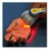
Laser Treatment - Low-Level: Low-level laser therapy (LLLT) is a medical treatment that applies low-level (low-power) lasers or light-emitting diodes (LEDs) to the surface or orifices of the body. Whereas "high-power" lasers are used in laser medicine to cut or destroy tissue, low-power lasers are claimed to relieve pain or to stimulate and enhance cell function.

The effects of LLLT appear to be limited to a specified set of wavelengths of laser, and administering LLLT below the dose range does not appear to be effective.

Despite a lack of consensus over its validity, some studies suggest that LLLT may be modestly effective, but in most cases no better than placebo, in relieving short-term pain for rheumatoid arthritis, osteoarthritis, acute and chronic neck pain, tendinopathy, and possibly chronic joint disorders. The evidence for LLLT being useful in the treatment of low back pain, dentistry and wound healing is unclear.

  • A comparative study of red and blue light-emitting diodes and low-level laser in regeneration of the transected sciatic nerve after an end to end neurorrhaphy in rabbits.

    facebook Share on Facebook
    Abstract Title:

    A comparative study of red and blue light-emitting diodes and low-level laser in regeneration of the transected sciatic nerve after an end to end neurorrhaphy in rabbits.

    Abstract Source:

    Lasers Med Sci. 2015 Sep 28. Epub 2015 Sep 28. PMID: 26415928

    Abstract Author(s):

    Mohammad Ashrafzadeh Takhtfooladi, Davood Sharifi

    Article Affiliation:

    Mohammad Ashrafzadeh Takhtfooladi

    Abstract:

    This study aimed at evaluating the effects of red and blue light-emitting diodes (LED) and low-level laser (LLL) on the regeneration of the transected sciatic nerve after an end-to-end neurorrhaphy in rabbits. Forty healthy mature male New Zealand rabbits were randomly assigned into four experimental groups: control, LLL (680 nm), red LED (650 nm), and blue LED (450 nm). All animals underwent the right sciatic nerve neurotmesis injury under general anesthesia and end-to-end anastomosis. The phototherapy was initiated on the first postoperative day and lasted for 14 consecutive days at the same time of the day. On the30th day post-surgery, the animals whose sciatic nerves were harvested for histopathological analysis were euthanized. The nerves were analyzed and quantified the following findings: Schwann cells, large myelinic axons, and neurons. In the LLL group, as compared to other groups, an increase in thenumber of all analyzed aspects was observed with significance level (P < 0.05). This finding suggests that postoperative LLL irradiation was able to accelerate and potentialize the peripheral nerve regeneration process in rabbits within 14 days of irradiation.

  • A randomized pilot study to assess the safety and the value of low-level laser therapy versus clonazepam in patients with burning mouth syndrome.

    facebook Share on Facebook
    Abstract Title:

    A randomized pilot study to assess the safety and the value of low-level laser therapy versus clonazepam in patients with burning mouth syndrome.

    Abstract Source:

    Lasers Med Sci. 2016 Feb 12. Epub 2016 Feb 12. PMID: 26873501

    Abstract Author(s):

    Paolo G Arduino, Adriana Cafaro, Marco Garrone, Alessio Gambino, Marco Cabras, Ercole Romagnoli, Roberto Broccoletti

    Article Affiliation:

    Paolo G Arduino

    Abstract:

    Comparison between low-level laser therapy (LLLT) and clonazepam for treating burning mouth syndrome (BMS) patients has never been documented; the aim of this study was to assess the effects of LLLT photobiomodulation versus medical therapy with clonazepam on BMS. Thirty-three patients (25 female, 8 male, mean age = 67.12) were randomly allocated to two different groups: the first one (group A, 18 patients) underwent two laser irradiation sessions weekly for 5 weeks, whereas the second one (group B, 15 patients) received topical clonazepam therapy [half a tablet (2 mg) in the mouth without swallowing for 3 min, three times a day for 21 days]. LLLT was delivered with a continuous wave 980-nm aluminum gallium arsenide (AlGaAs) diode laser and the output of 300 mW, delivering a Fluence of 10 J/cm(2), using a"spot technique,"with an average power density of about 1 W/cm(2). The laser probe was held perpendicularly at a distance of about 2 mm from the mucosa. Visual analogue scale (VAS), McGill Pain Questionnaire, present pain intensity (PPI), and Oral Health Impact Profile (OHIP-49) assessed sensation of pain. Hospital Anxiety and Depression Scale and Geriatric Depression Scale assessed levels of anxiety and depression. Twelve weeks after the end of treatment, patients treated with LLLT experienced a decrease in pain sensation reported for all the parameters analyzed: VAS (P = 0.004), McGill Pain Questionnaire (P = 0.002), PPI (P = 0.002),and OHIP-49 (P = 0.010). The group treated with clonazepam had less favorable results for VAS (P = 0.33), McGill Pain Questionnaire (P = 0.005), PPI (P = 0.013), and OHIP-49 (P = 0.25). Levels of anxiety and depression did not change statistically in any groups (P > 0.05). Comparing the two groups, LLLT appeared to be superior in improving pain perception, but statistically only at 8 weeks after the end of the protocol proposed (P = 0.026). Based on this preliminary trial, LLLT is capable of reducing the symptoms of patients with BMS with a constant andlong-lasting effect, experienced since the end of the first applications.

  • Adjunct Use of Low-Level Laser Therapy on the Treatment of Necrotizing Ulcerative Gingivitis: A Case Report. 📎

    facebook Share on Facebook
    Abstract Title:

    Adjunct Use of Low-Level Laser Therapy on the Treatment of Necrotizing Ulcerative Gingivitis: A Case Report.

    Abstract Source:

    J Lasers Med Sci. 2018 ;9(1):73-75. Epub 2017 Dec 26. PMID: 29399316

    Abstract Author(s):

    Seda Sevinç Özberk, Hasan Gündoğar, Süleyman Ziya Şenyurt, Kamile Erciyas

    Article Affiliation:

    Seda Sevinç Özberk

    Abstract:

    Necrotizing ulcerative gingivitis (NUG) is a microbial disease of the gingiva in the context of an impaired host response. This form of gingivitis is relatively rare. NUG is an infection characterized by gingival necrosis presenting as"punched-out"papillae, spontaneous bleeding, pain, oral malodor, and pseudomembrane formation. The primary predisposing factors are bacterial plaque and an inadequate diet, but smoking and psychological stress may also affect the disease severity. NUG is associated with a characteristic bacterial flora, which includes fusiform bacteria, spirochetes, and Prevotella intermedia. Conventional treatment includes control of both the bacterial plaque and the secondary factors, as well as topical or systemic treatment biostimulative effect on wound healing, pain control, and inflammatory processes. Patients with NUG were treated using adjunct use of a diode laser (980 nm) for the control of pain and to accelerate the wound healing at day 2. 3. 5. 9, energy density was 9 J/cm. After treatment, the patients' quality of life improved faster than with conventional treatment. These results suggest that low-level laser therapy (LLLT) is an effective treatment for the reduction of pain levels and healing times. As a result, our case report shows that LLTT has a positive effect in relieving the symptoms of NUG.

  • Anti-inflammatory effects of low-level laser therapy on human periodontal ligament cells: in vitro study. 📎

    facebook Share on Facebook
    Abstract Title:

    Anti-inflammatory effects of low-level laser therapy on human periodontal ligament cells: in vitro study.

    Abstract Source:

    Lasers Med Sci. 2017 Nov 7. Epub 2017 Nov 7. PMID: 29116611

    Abstract Author(s):

    Ji-Hua Lee, Min-Hsuan Chiang, Ping-Ho Chen, Mei-Ling Ho, Huey-Er Lee, Yan-Hsiung Wang

    Article Affiliation:

    Ji-Hua Lee

    Abstract:

    Periodontal disease is a chronic inflammatory disease that is commonly treated with surgical and nonsurgical techniques. However, both approaches have limitations. Low-level laser therapy (LLLT) has been widely applied in reducing inflammatory reactions, and research indicates that LLLT induces an anti-inflammatory effect that may enhance periodontal disease therapy. The purpose of this study was to investigate the anti-inflammatory effect of LLLT on human periodontal ligament cells (hPDLCs) in an inflammatory environment and aimed to determine the possible mechanism of action. Cells were cultured and treated with or without lipopolysaccharide (LPS) from Porphryromonas gingivalis or Escherichia coli, followed by irradiation with a gallium-aluminum-arsenide (GaAlAs) laser (660 nm) at an energy density of 8 J/cm2. Quantitative real-time polymerase chain reactions were used to assess the expression of pro-inflammatory genes, including tumor necrosis factor-α (TNF-α), interleukin (IL)-1β, IL-6, and IL-8. The dual-luciferase reporter assay was used to examine nuclear factor-κB (NF-κB) transcriptional activity. An enzyme-linked immunosorbent assay was used to monitor the concentration of intracellular cyclic adenosine monophosphate (cAMP). Both LPS treatments significantly induced the mRNA expression of pro-inflammatory cytokines. However, LLLT inhibited the LPS-induced pro-inflammatory cytokine expression and elevated intracellular levels of cAMP. The LLLT inhibitory effect may function by downregulating NF-κB transcriptional activity and by increasing the intracellular levels of cAMP. LLLT might inhibit LPS-induced inflammation in hPDLCs through cAMP/NF-κB regulation. These results should be further studied to improve periodontal therapy.

  • Chapter 25: Phototherapy in peripheral nerve injury: effects on muscle preservation and nerve regeneration.

    facebook Share on Facebook
    Abstract Title:

    Chapter 25: Phototherapy in peripheral nerve injury: effects on muscle preservation and nerve regeneration.

    Abstract Source:

    Int Rev Neurobiol. 2009;87:445-64. PMID: 19682654

    Abstract Author(s):

    Shimon Rochkind, Stefano Geuna, Asher Shainberg

    Article Affiliation:

    Division of Peripheral Nerve Reconstruction, Department of Neurosurgery, Tel Aviv Sourasky Medical Center, Tel Aviv University, Israel.

    Abstract:

    Posttraumatic nerve repair and prevention of muscle atrophy represent a major challenge of restorative medicine. Considerable interest exists in the potential therapeutic value of laser phototherapy for restoring or temporarily preventing denervated muscle atrophy as well as enhancing regeneration of severely injured peripheral nerves. Low-power laser irradiation (laser phototherapy) was applied for treatment of rat denervated muscle in order to estimate biochemical transformation on cellular and tissue levels, as well as on rat sciatic nerve model after crush injury, direct or side-to-end anastomosis, and neurotube reconstruction. Nerve cells' growth and axonal sprouting were investigated in embryonic rat brain cultures. The animal outcome allowed clinical double-blind, placebo-controlled randomized study that measured the effectiveness of 780-nm laser phototherapy on patients suffering from incomplete peripheral nerve injuries for 6 months up to several years. In denervated muscles, animal study suggests that the function of denervated muscles can be partially preserved by temporary prevention of denervation-induced biochemical changes. The function of denervated muscles can be restored, not completely but to a very substantial degree, by laser treatment initiated at the earliest possible stage post injury. In peripheral nerve injury, laser phototherapy has an immediate protective effect. It maintains functional activity of the injured nerve for a long period, decreases scar tissue formation at the injury site, decreases degeneration in corresponding motor neurons of the spinal cord, and significantly increases axonal growth and myelinization. In cell cultures, laser irradiation accelerates migration, nerve cell growth, and fiber sprouting. In a pilot, clinical, double-blind, placebo-controlled randomized study in patients with incomplete long-term peripheral nerve injury, 780-nm laser irradiation can progressively improve peripheral nerve function, which leads to significant functional recovery. A 780-nm laser phototherapy temporarily preserves the function of a denervated muscle, and accelerates and enhances axonal growth and regeneration after peripheral nerve injury or reconstructive procedures. Laser activation of nerve cells, their growth, and axonal sprouting can be considered as potential treatment for neural injury. Animal and clinical studies show the promoting action of phototherapy on peripheral nerve regeneration, which makes it possible to suggest that the time for broader clinical trials has come.

  • Chiropractic manipulative therapy and low-level laser therapy in the management of cervical facet dysfunction: a randomized controlled study.

    facebook Share on Facebook
    Abstract Title:

    Chiropractic manipulative therapy and low-level laser therapy in the management of cervical facet dysfunction: a randomized controlled study.

    Abstract Source:

    J Manipulative Physiol Ther. 2011 Mar-Apr;34(3):153-63. PMID: 21492750

    Abstract Author(s):

    Lindie Saayman, Caroline Hay, Heidi Abrahamse

    Article Affiliation:

    Laser Research Centre, Faculty of Health Sciences, University of Johannesburg, Doornfontein, South Africa.

    Abstract:

    PURPOSE:The aim of this study was to determine the short-term effect of chiropractic joint manipulation therapy (CMT) and low-level laser therapy (LLLT) on pain and range of motion in the management of cervical facet dysfunction.

    METHODS:Sixty ambulatory women between the ages of 18 and 40 years with cervical facet joint pain of more than 30-day duration and normal neurologic examination were randomized to receive 1 of 3 treatment options: (1) CMT of the cervical spine, (2) LLLT applied to the cervical facet joints, or (3) a combination of CMT and LLLT. Each participant received 6 treatments in 3 weeks. The main outcome measures were as follows: the Numerical Pain Rating Scale, Neck Disability Index, Cervical Range of Motion Instrument, and Baseline Digital Inclinometer. Measurements were taken during weeks 1 (baseline), 2, 3, and 4.

    RESULTS:No differences existed between the 3 groups at baseline. A significant difference was seen between groups 1 (CMT) and 2 (LLLT) for cervical flexion, between groups 1 (CMT) and 3 (CMT + LLLT) for cervical flexion and rotation, and between groups 2 (LLLT) and 3 (CMT + LLLT) for pain disability in everyday life, lateral flexion, and rotation.

    CONCLUSION:All 3 groups showed improvement in the primary and secondary outcomes. A combination of CMT and LLLT was more effective than either of the 2 on their own. Both therapies are indicated as potentially beneficial treatments for cervical facet dysfunction. Further studies are needed to explore optimal treatment procedures for CMT and LLLT and the possible mechanism of interaction between therapies.

  • Combined therapeutic effects of low power laser (980nm) and CoQ10 on Neuropathic Pain in adult male rat. 📎

    facebook Share on Facebook
    Abstract Title:

    Combined therapeutic effects of low power laser (980nm) and CoQ10 on Neuropathic Pain in adult male rat.

    Abstract Source:

    Med J Islam Repub Iran. 2014 ;28:58. Epub 2014 Jul 13. PMID: 25405124

    Abstract Author(s):

    Seyed Behnamedin Jameie, Masoumeh Masoumipoor, Atousa Janzadeh, Farinaz Nasirinezhad, Mahdieh Kerdari, Maryam Soleimani

    Article Affiliation:

    Seyed Behnamedin Jameie

    Abstract:

    BACKGROUND:Neuropathic pain (NP) is one of the most suffering medical conditions that often fail to respond to certain pain therapy. Although its exact etiology is still unknown the role of reactive oxygen species (ROS) and oxidative stress were explored by many researchers. Neuropathies either central or peripheral lead to painful condition as well as social and economic isolation, thus various therapies were used to treat or reduce the pain. Laser therapy and antioxidant drugs have separately considered as treatment for NP, but the combination of them have not been used yet. In order to study the combination effects of Low Level Laser Therapy (LLLT) and Coenzyme Q10 (CoQ10) the present study was designed.

    METHODS:Sixty adult male rats (230-320g) were used in this experimental study that divided into six groups (n=10). Chronic constriction injury (CCI) was used to induce neuropathic pain. The CoQ10 or vehicle, a low level laser of 980nm was used for two consecutive weeks. Thermal and mechanical paw withdrawal thresholds were assessed before and after surgery on 7(th) and 14(th) days.

    RESULTS:As we expected CCI decreased the pain threshold, whereas CoQ10 administration for two weeks increased mechanical and thermal threshold. The same results obtained for laser therapy using the CCI animals. Combination of laser 980nm with CoQ10 also showed significant differences in CCI animals.

    CONCLUSION:Based on our findings the combination of CoQ10 with LLLT showed better effects than each one alone. In this regard we believe that there might be cellular and molecular synergism in simultaneous use of CoQ10 and LLLT on pain relief.

  • Comparison of laser and ozone treatments on oral mucositis in an experimental model.

    facebook Share on Facebook
    Abstract Title:

    Comparison of laser and ozone treatments on oral mucositis in an experimental model.

    Abstract Source:

    Lasers Med Sci. 2017 Apr ;32(3):673-677. Epub 2017 Feb 11. PMID: 28190112

    Abstract Author(s):

    Suzan Bayer, Hakki Oguz Kazancioglu, Ahmet Hüseyin Acar, Nihat Demirtas, Nur Ozten Kandas

    Article Affiliation:

    Suzan Bayer

    Abstract:

    Oral mucositis (OM) induces severe pain and limits fundamental life behaviors such as eating, drinking, and talking for patients receiving chemotherapy or radiotherapy. In addition, through opportunistic microorganisms, OM frequently leads to systemic infection which then leads to prolonged hospitalization. Severe lesions often adversely affect curative effects in cancer cases. Therefore, the control of OM is important for oral health quality of life and prognosis. Low-level laser therapy (LLLT) and ozone may be useful to accelerate wound healing. In this study, 24 Sprague-Dawley rats were divided into three groups as control, ozone, and laser groups. All groups received 5-fluorouracil intraperitoneally and trauma to the mouth pouch with a needle. After the formation of OM in the mouth, the control group had no treatment; the ozone group was administered ozone, and the laser group, LLLT. Then, all groups were sacrificed and basic fibroblast growth factor (bFGF), transforming growth factor (TGF-β), and platelet-derived growth factor (PDGF) were evaluated in all groups. LLLT was determined to be statistically significantly more effective than ozone on FGF and PDGF. However, in respect of TGF-β, no statistically significant difference was observed between the groups. In conclusion, withinthe limitations of this study, LLLT is more effective than ozone. However, further studies on this subject are required.

  • Comparison of Photobiomodulation and Anti-Inflammatory Drugs on Tissue Repair on Collagenase-Induced Achilles Tendon Inflammation in Rats.

    facebook Share on Facebook
    Abstract Title:

    Comparison of Photobiomodulation and Anti-Inflammatory Drugs on Tissue Repair on Collagenase-Induced Achilles Tendon Inflammation in Rats.

    Abstract Source:

    Photomed Laser Surg. 2017 Dec 21. Epub 2017 Dec 21. PMID: 29265910

    Abstract Author(s):

    Ingvill Fjell Naterstad, Rafael Paolo Rossi, Rodrigo Labat Marcos, Nivaldo Antonio Parizzoto, Lucio Frigo, Jón Joensen, Patrícia Sardinha Leonardo Lopes Martins, Jan Magnus Bjordal, Rodrigo Alvaro Brandão Lopes-Martins

    Article Affiliation:

    Ingvill Fjell Naterstad

    Abstract:

    BACKGROUND:Tendinopathy is characterized by pain, edema, and structural changes in tendon tissue.

    OBJECTIVE:In this animal study we decided to compare the short- and medium-term effects of low-level laser therapy (LLLT), dexamethasone, and diclofenac on inflammation and tendon tissue repair in collagenase-induced tendinitis.

    MATERIALS AND METHODS:Two hundred five female Wistar rats were randomly divided into five groups. Animals in the control group were given a saline injection and the experimental groups received a collagenase injection (100 μg/tendon) in the peritendinous Achilles and received no treatment, LLLT (3 J, 810 nm, 100 mW), diclofenac (1.1 mg/kg), or dexamethasone (0.02 mg/kg). Histological analyses were performed at 10 time points up to 60 days (n = 5/group each time point), and included an assessment of the severity of inflammation, collagen fiber content, and organization.

    RESULTS:Collagenase injection induced a severe inflammatory reaction with significant reduction in collagen content for 48 h, and disorientation of collagen fibers lasting between 14 and 21 days. Diclofenac and dexamethasone reduced inflammatory signs during the first 2 days, although there was prolongation of the inflammatory phase and slower normalization of tendon quality, particularly in the dexamethasone group.LLLT prevented hemorrhage, reduced inflammation severity, and preserved tendon morphology compared with the other groups.

    CONCLUSIONS:LLLT showed a significant superiority over commonly used anti-inflammatory pharmaceutical agents in acute collagenase-induced tendinitis.

  • Contribution of photodynamic therapy in wound healing: A systematic review.

    facebook Share on Facebook
    Abstract Title:

    Contribution of photodynamic therapy in wound healing: A systematic review.

    Abstract Source:

    Photodiagnosis Photodyn Ther. 2018 Mar ;21:294-305. Epub 2017 Dec 28. PMID: 29289704

    Abstract Author(s):

    Vanessa Nesi-Reis, Daniele Stéfanie Sara Lopes Lera-Nonose, Jully Oyama, Marielle Priscila Paula Silva-Lalucci, Izabel Galhardo Demarchi, Sandra Mara Alessi Aristides, Jorge Juarez Vieira Teixeira, Thaís Gomes Verzignassi Silveira, Maria Valdrinez Campana Lonardoni

    Article Affiliation:

    Vanessa Nesi-Reis

    Abstract:

    OBJECTIVE:We researched articles that used photodynamic therapy (PDT) for skin wound healing in humans.

    METHODS:The systematic review was conducted through scientific articles that investigated the action of PDT on wound healing in humans, published from July 2005 to March 2017, in the data bases PubMed and LILACS.

    RESULTS:The main types of wound described in selected articles in this review were chronic ulcer and non-melanoma skin cancer. For accomplishing the PDT, second generation of photosensitizing agents with laser or light emitting diode were used. The studies demonstrated that PDT contribute in several ways to the wound healing process: leading to cellular death; reducing or increasing inflammation; stimulating fibroblasts proliferation and, consequently, of collagen and elastin; raising transforming growth factor beta and metalloproteinases. Based on this, PDT provided good results in wound healing process, acting in several steps and accelerating tissue repair.

    CONCLUSIONS:PDT improved healing in many wound models in humans, revealing itself as a promising therapeutic modality for stimulating wound healing and remodelling.

  • Cultured epithelial cells response to phototherapy with low intensity laser.

    facebook Share on Facebook
    Abstract Title:

    Cultured epithelial cells response to phototherapy with low intensity laser.

    Abstract Source:

    Lasers Surg Med. 2007 Apr;39(4):365-72. PMID: 17457843

    Abstract Author(s):

    Fernanda P Eduardo, Dolores U Mehnert, Telma A Monezi, Denise M Zezell, Mark M Schubert, Carlos P Eduardo, Márcia M Marques

    Article Affiliation:

    Department of Oral Diagnosis, School of Dentistry, University of São Paulo, São Paulo, Brazil.

    Abstract:

    BACKGROUND AND OBJECTIVES:Little is known about the intracellular response of epithelial cells to phototherapy. The aim of this in vitro study was to analyze the effect of phototherapy with low-energy lasers with different wavelengths and powers on cultured epithelial cell growth under different nutritional conditions.

    STUDY DESIGN/MATERIALS AND METHODS:Epithelial cell cultures (Vero cell line) grown in nutritional deficit in culture medium supplemented with 2% fetal bovine serum (FBS) were irradiated with low-energy laser from one to three times with a GaAlAs laser (660 nm) and InGaAlP (780 nm), 40 and 70 mW, respectively, with 3 or 5 J/cm2. Cell growth was indirectly assessed by measuring the cell mitochondrial activity.

    RESULTS:Nonirradiated cell cultures grown in nutritional regular medium supplemented with 10% FBS produced higher cell growth than all cultures grown in nutritional deficit irradiated or not. The overall cell growth of cultures grown under nutritionally deficit conditions was significantly improved especially when irradiated with 780 nm for three times.

    CONCLUSIONS:Phototherapy with the laser parameters tested increases epithelial cell growth rate for cells stressed by growth under nutritionally deficient states. This cell growth improvement is directly proportional to the number of irradiations; however, was not enough to reach the full cell growth potential rate of Vero epithelial cell line observed when growing under nutritional regular condition.

  • Effect of low energy light irradiation by light emitting diode on U937 cells.

    facebook Share on Facebook
    Abstract Title:

    Effect of low energy light irradiation by light emitting diode on U937 cells.

    Abstract Source:

    J Biol Regul Homeost Agents. 2016 Oct-Dec;30(4):997-1007. PMID: 28078845

    Abstract Author(s):

    G Spoto, V De Iuliis, M Petrini, V Flati, J Di Gregorio, D Vitale, M Caruso, V Dadorante, M Ciarmoli, I Robuffo, S Martinotti, E Toniato

    Article Affiliation:

    G Spoto

    Abstract:

    Photobiomodulation (PBM) can induce a set of different biological modulators either in vitro or in vivo. Experimental evidence has highlighted the role of light effects on the mechanisms related to inflammation, apoptosis and autophagy. The goal of this project was the evaluation of PBM on U937, an established cell line of histiocytic lymphoma origin. Several aspects of modulation of proinflammatory pathways were analyzed and autophagic and proapoptotic mechanisms related to low laser light exposure of cells were studied. As a source of low energy light emission, we used an NIR-LED device, characterized by an 880 nm-wavelength as light source. Flow cytometry analysis was performed on supernatants of controls and treated U937 cells to detect inflammatory cytokine levels. In order to evaluate NF-kB and caspase3 expressions, Western blot analysis was performed according to standard procedures. In this report, we show the effect of PBM on a monocyte/macrophage established tumor cell line (U-937). We demonstrate that LED exposure, in the presence or absence of lipopolysaccharide (LPS), activates cell degranulation, increased expression of Interleukin-8 (IL-8) and modulation of beta galactosidase activity. Evidence shows that the well-known pro-inflammatory nuclear factor kappa-light-chain-enhancer of activated B cells (NF-kB) and the apoptotic marker (caspase3/cleaved-caspase3 ratio) are up-regulated in response to a proinflammatory biochemical pathway.

  • Effect of low level laser therapy in rheumatoid arthritis patients with carpal tunnel syndrome.

    facebook Share on Facebook
    Abstract Title:

    Effect of low level laser therapy in rheumatoid arthritis patients with carpal tunnel syndrome.

    Abstract Source:

    Swiss Med Wkly. 2007 Jun 16 ;137(23-24):347-52. PMID: 17629805

    Abstract Author(s):

    Ayse Ekim, Onur Armagan, Funda Tascioglu, Cengiz Oner, Meric Colak

    Article Affiliation:

    Ayse Ekim

    Abstract:

    OBJECTIVE:the aim of the present study was to evaluate the efficacy of low level laser therapy (LLLT) in patients with rheumatoid arthritis (RA) with carpal tunnel syndrome (CTS).

    MATERIAL AND METHODS:a total of 19 patients with the diagnosis of CTS in 19 hands were included and randomly assigned to two treatment groups; LLLT (Group 1) (10 hands) with dosage 1.5 J/ per point and placebo laser therapy group (Group 2) (9 hands). A Galium-Aluminum-Arsenide diode laser device was used as a source of low power laser with a power output of 50 mW and wavelength of 780 nm. All treatments were applied once a day on week days for a total period of 10 days. Clinical assessments were performed at baseline, at the end of the treatment and at month 3. Tinel and Phalen signs were tested in all patients. Patients were evaluated for such clinical parameters as functional status scale (FSS), visual analogue scale (VAS), symptom severity scale (SSS) and grip-strength. However, electrophysiological examination was performed on all hands. Results were given with descriptive statistics and confidence intervals between group means at 3 months adjusted for outcome at baseline and for the difference between unadjusted group proportions.

    RESULTS:clinical and electrophysiological parameters were similar at baseline in both groups. Improvements were significantly more pronounced in the LLLT group than placebo group. A comparison between groups showed significant improvements in pain score and functional status scale score. Group mean differences at 3 months adjusted at baseline were found to be statistically significant for pain score and functional status scale score. The 95% significant confidence intervals were [-15 - (-5)] and [-5 - (-2)] respectively. There were no statistically significant differences in other clinical and electrophysiological parameters between groups at 3 months.

    CONCLUSIONS:our study results indicate that LLLT and placebo laser therapy seems to be effective for pain and hand function in CTS. We, therefore, suggest that LLLT may be used as a good alternative treatment method in CTS patients with RA.

  • Effect of Low-Level Laser Therapy on Bone Regeneration During Osseointegration and Bone Graft.

    facebook Share on Facebook
    Abstract Title:

    Effect of Low-Level Laser Therapy on Bone Regeneration During Osseointegration and Bone Graft.

    Abstract Source:

    Photomed Laser Surg. 2017 Jul 21. Epub 2017 Jul 21. PMID: 28742438

    Abstract Author(s):

    Randa Zein, Wayne Selting, Stefano Benedicenti

    Article Affiliation:

    Randa Zein

    Abstract:

    BACKGROUND:The effect of low-level laser therapy (LLLT) on bone regeneration during osseointegration and bone graft is very controversial. Despite many positive reports of in vitro and in vivo studies and more than 50 randomized clinical trials claiming a positive effect of photobiomodulation (PBM), many reports found no significant effect of lasers.

    OBJECTIVE:The aim of this study was to evaluate studies correlating PBM and bone regeneration and to assesses parameters that produce positive results based on dose and output power used.

    MATERIALS AND METHODS:Four electronic databases were used: PubMed, Springer, Google Scholar, and Cochrane.

    RESULTS:The research yielded 230 articles. The full texts of all articles were evaluated and scored using eligibility criteria adapted from Cericato et al. After evaluation, only 19 articles met the inclusion criteria.

    CONCLUSIONS:A positive effect of low-level laser energy on bone regeneration within a certain relationship between dose and output power was found. LLLT stimulates cellular metabolism, increasing protein synthesis and subsequent bone regeneration. A high dose combined with low power or a low dose combined with high power appears to produce a positive effect.

  • Effect of low-level laser therapy on the healing process of donor site in patients with grade 3 burn ulcer after skin graft surgery (a randomized clinical trial).

    facebook Share on Facebook
    Abstract Title:

    Effect of low-level laser therapy on the healing process of donor site in patients with grade 3 burn ulcer after skin graft surgery (a randomized clinical trial).

    Abstract Source:

    Lasers Med Sci. 2018 Apr ;33(3):603-607. Epub 2018 Jan 24. PMID: 29368069

    Abstract Author(s):

    Reza Vaghardoost, Mahnoush Momeni, Nooshafarin Kazemikhoo, Soheila Mokmeli, Mostafa Dahmardehei, Fereshteh Ansari, Mohammad Ali Nilforoushzadeh, Parisa Sabr Joo, Sara Mey Abadi, Soheila Naderi Gharagheshlagh, Saeed Sassani

    Article Affiliation:

    Reza Vaghardoost

    Abstract:

    Skin graft is a standard therapeutic technique in patients with deep ulcers, but managing donor site after grafting is very important. Although several modern dressings are available to enhance the comfort of donor site, using techniques that accelerate wound healing may enhance patient satisfaction. Low-level laser therapy (LLLT) has been used in several medical fields, including healing of diabetic, surgical, and pressure ulcers, but there is not any report of using this method for healing of donor site in burn patients. The protocols and informed consent were reviewed according to Medical Ethics Board of Shahid Beheshti University of Medical Sciences (IR.SBMU.REC.1394.363) and Iranian Registry of Clinical Trials (IRCT2016020226069N2). Eighteen donor sites in 11 patients with grade 3 burn ulcer were selected. Donor areas were divided into 2 parts, for laser irradiation and control randomly. Laser area was irradiated by a red, 655-nm laser light, 150 mW, 2 J/cm, on days 0 (immediately after surgery), 3, 5, and 7. Dressing and other therapeutic care for both sites were the same. The patients and the person who analyzed the results were blinded. The size of donor site reduced in both groups during the 7-day study period (P < 0.01) and this reduction was significantly greater in the laser group (P = 0.01). In the present study, for the first time, we evaluate the effects of LLLT on the healing process of donor site in burn patients. The results showed that local irradiation of red laser accelerates wound healing process significantly.

  • Effect of photobiomodulation and exercise on early remodeling of the Achilles tendon in streptozotocin-induced diabetic rats📎

    facebook Share on Facebook
    Abstract Title:

    Effect of photobiomodulation and exercise on early remodeling of the Achilles tendon in streptozotocin-induced diabetic rats.

    Abstract Source:

    PLoS One. 2019 ;14(2):e0211643. Epub 2019 Feb 4. PMID: 30716140

    Abstract Author(s):

    Anderson Rodrigues de Oliveira, Flávio Santos da Silva, Raul Hernandes Bortolin, Dáfiny Emanuele da Silva Marques, Gracielle Vieira Ramos, Rita C Marqueti, Naisandra Bezerra da Silva, Karina Carla de Paula Medeiros, Márcio Assolin Corrêa, João Paulo Matos Santos Lima, Adriana Augusto de Rezende, Paul W Ackermann, Bento J Abreu, Wouber Hérickson de Brito Vieira

    Article Affiliation:

    Anderson Rodrigues de Oliveira

    Abstract:

    The aim of this study was to compare the treatment effects of laser photobiomodulation (LPBM) therapy and aerobic exercise on the biomechanical properties, tissue morphology and the expression of tendon matrix molecules during early remodeling of Achilles tendon (AT) injury in diabetic rats. Animals were randomly assigned to five groups: injured non diabetic (I, n = 15), injured diabetic (ID, n = 15), injured diabetic plus LPBM (IDL, n = 16), injured diabetic plus aerobic exercise (IDE, n = 16) and injured diabetic plus aerobic exercise and LPBM (IDEAL, n = 17). Type 1 diabetes was induced via a single intravenous injection of Streptozotocin at a dose of 40 mg/kg. A partial tenotomy was performed in the right AT. LPBM was performed with an indium-gallium-aluminum-phosphide 660 nm 10 mW laser device (spot size 0.04 cm2, power density 250 mW/cm2, irradiation duration 16 s, energy 0.16 J, energy density 4 J/cm2) on alternate days for a total of 9 sessions over 3 weeks (total energy 1.44 J), using a stationary contact technique to a single point over the dorsal aspect of the AT. Moderate aerobic exercise was performed on a motorized treadmill (velocity 9 m/min for 60 minutes). At 3 weeks post-injury, biomechanical analyzes as well as assessment of fibroblast number and orientation were performed. Collagen 1 (Col1) and 3 (Col3) and matrix metalloproteinases (MMPs) -3 and 13 protein distributions were studied by immunohistochemistry; while Col1 and Col3 and MMP-2 and 9 gene expression were assessed by quantitative RT-PCR (qRT-PCR). IDEAL exhibited significant increases in several biomechanical parameters in comparison to the other groups. Moreover, IDEAL presented stronger Col1 immunoreactivity when compared to ID, and weaker Col3 immunoreactivity than IDE. Both IDL and IDEAL demonstrated weaker expression of MMP-3 in comparison to I, while IDL presented no expression of MMP-13 when compared to ID. ID, IDL and IDE showed an increased number of fibroblasts in comparison to I, while IDEAL decreased the number of these cells in comparison to ID and IDE. IDL and IDEAL groups exhibited decreased angular dispersion among the fibroblasts when compared to I. The gene expression results showed that IDE demonstrated a downregulation in Col1 mRNA expression in comparison to I and ID. IDEAL demonstrated upregulation of Col1 mRNA expression when compared to IDL or IDE alone and increased MMP-2 expression when compared to IDL and IDE. MMP-9 expression was upregulated in IDEAL when compared to I, IDL and IDE. Our results suggest a beneficial interaction of combining both treatment strategies i.e., aerobic exercise and LPBM, on the biomechanical properties, tissue morphology and the expression of matrix molecules in diabetic tendons.

  • Effect of red and near-infrared wavelengths on low-level laser (light) therapy-induced healing of partial-thickness dermal abrasion in mice. 📎

    facebook Share on Facebook
    Abstract Title:

    Effect of red and near-infrared wavelengths on low-level laser (light) therapy-induced healing of partial-thickness dermal abrasion in mice.

    Abstract Source:

    Lasers Med Sci. 2014 Jan ;29(1):257-65. Epub 2013 Apr 26. PMID: 23619627

    Abstract Author(s):

    Asheesh Gupta, Tianhong Dai, Michael R Hamblin

    Article Affiliation:

    Asheesh Gupta

    Abstract:

    Low-level laser (light) therapy (LLLT) promotes wound healing, reduces pain and inflammation, and prevents tissue death. Studies have explored the effects of various radiant exposures on the effect of LLLT; however, studies of wavelength dependency in in vivo models are less common. In the present study, the healing effects of LLLT mediated by different wavelengths of light in the red and near-infrared (NIR) wavelength regions (635, 730, 810, and 980 nm) delivered at constant fluence (4 J/cm(2)) and fluence rate (10 mW/cm(2)) were evaluated in a mouse model of partial-thickness dermal abrasion. Wavelengths of 635 and 810 nm were found to be effective in promoting the healing of dermal abrasions. However, treatment using 730- and 980-nm wavelengths showed no sign of stimulated healing. Healing was maximally augmented in mice treated with an 810-nm wavelength, as evidenced by significant wound area reduction (p<0.05), enhanced collagen accumulation, and complete re-epithelialization as compared to other wavelengths and non-illuminated controls. Significant acceleration of re-epithelialization and cellular proliferation revealed by immunofluorescence staining for cytokeratin-14 and proliferating cell nuclear antigen (p<0.05) was evident in the 810-nm wavelength compared with other groups. Photobiomodulation mediated by red (635 nm) and NIR (810 nm) light suggests that the biological response of the wound tissue depends on the wavelength employed. The effectiveness of 810-nm wavelength agrees with previous publications and, together with the partial effectiveness of 635 nm and the ineffectiveness of 730 and 980 nm wavelengths, can be explained by the absorption spectrum of cytochrome c oxidase, the candidate mitochondrial chromophore in LLLT.

We use cookies on our website. Some of them are essential for the operation of the site, while others help us to improve this site and the user experience (tracking cookies). You can decide for yourself whether you want to allow cookies or not. Please note that if you reject them, you may not be able to use all the functionalities of the site.